STIM1 signaling pathway
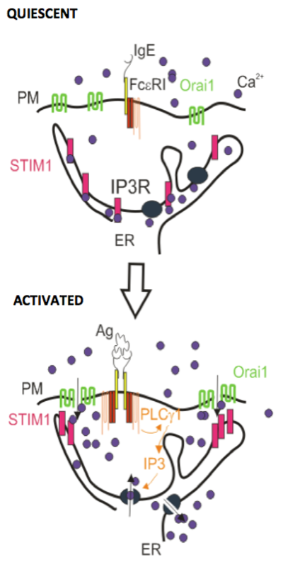
The stromal interaction molecule 1 (STIM1) is a protein embedded predominantly in endoplasmic reticulum (ER) membrane via its transmembrane domain. It is a key regulator of calcium release activated calcium (CRAC) channel responsible for store operated calcium entry (SOCE) formerly known as Ca2+ capacitative entry. The function of STIM1 remained elusive until 2005 when two independent studies based on large-scale siRNA screens identified STIM1 as a regulator of SOCE (1, 2). A year later Orai1 was described as the crucial CRAC channel and thus the basic molecular mechanism of enigmatic SOCE was clarified (3-5). Orai1 together with Orai2 and 3 belong to the small family of conserved tetraspan proteins nonrelated to other channel families (6). STIM1 has a paralogue protein STIM2 (7). It has been reported that ectopicly expressed STIM2 can partially restore SOCE (8), and is probably involved in regulation of resting Ca2+ levels in ER (9). These SOCE activating proteins are ubiquitiously expressed; their pivotal role for calcium uptake was confirmed in many cell types including mast cells and basophils (10-12). STIM1 is capable of monitoring Ca2+ store levels via its conserved Ca2+ binding domain (EF hand) orientated in ER and responding to calcium depletion by aggregation. This process triggers a conformational change of the cytosolic part of STIM1 that releases highly conserved CRAC activation domain (CAD) also known as the STIM1 Orai1 activating region (SOAR) (13, 14). Puncta of oligomerized STIM1 are concentrated in a close vicinity to the plasma membrane (PM) where CAD can induce the formation of CRAC channels composed of Orais tetramers or other proteins, such as the TRPC channels (15). Some experimental data indicate that a small pool of STIM1 is present in PM and thus controls the arachidonic-acid-regulated Ca2+-selective channels (16). A study on mast cells proved that in ER-PM junctions STIM1 first bound to PIP2 in liquid ordered domains at the PM, followed by redistribution of Orai1 from liquid disordered regions of the PM to its association with STIM1 in liquid ordered lipid domains (17). An actin binding splice variant of STIM1, the STIM1L, was identified in some tissues forming permanent clusters colocalizing with Orai1 in ER-PM junctions and thus capable of immediatelly activating SOCE (18). The formation of STIM1 and Orai1 complex is regulated by CRACR2A protein which directly interacts with Orai1 and STIM1 and is important for their clustering (19). Dissociation of the CRAC channel components is Ca2+ dependent and two modes of negative feedback regulation have benn described. Fast inactivation depends mainly on STIM1 and calmodulin binding to Orai1 (20) and a slow Ca2+-dependent mechanism is based on a single ER membrane spanning protein SARAF that facilitates the disaggregation of STIMs molecules to efficiently turn off Orais when ER Ca2+ stores are refilled to basal levels (21). STIM1 is also able to interact with microtubule +TIP protein end binding 1 (EB1) and tracks its comet-like behavior (22). However, depletion of EB1 or inhibition of microtubule dynamics by taxol had no significant effect on SOCE in HeLa cells (22) but STIM1 mRNA silencing prevented changes in microtubule dynamics in thapsigargin-activated mast cells (23).
STIM1 in mast cells
Like in other nonexcitable cells, SOCE in mast cells is the major Ca2+ entry pathway. STIM1 aggregation is induced not only by crosslinking of the high affinity immunoglobulin E receptor (FcεRI) with antigen or dimerization of the stem cell factor receptor (c-Kit), but also by other pathways capable of activating inositol trisphosphate receptor that is responsible for depletion of Ca2+ stores. Antigen-mediated release of Ca2+ from intracellular stores followed by activation of SOCE is sufficient to induce mast cell degranulation, activate cell motility, chemotaxis, cytokine synthesis and produce arachidonic acid metabolites (24). Mice lacking STIM1 or Orai1 exhibit increased perinatal lethality and the growth of surviving mice is markedly retarded (12, 25). Mast cells derived from such knock out mice show defective degranulation and cytokine secretion (10, 12).
Human channelopaties; SOCE as a therapeutical targets
Patients with geneticaly inherited STIM1 or Orai1 disfunctions suffer from severe combined immunodeficiency syndroms characterized by normal T cell development but strongly impaired T cell activation (26). In vivo anaphylaxis studies on mice deficient in STIM1 or Orai1 indicate that mast cell activation is also strongly damaged (10, 12). Obviously, inhibitors of SOCE could serve as potential therapeutic agents for allergy and other deseases (27).
References:
| 1. | Liou, J., Kim, M. L., Do, H. W., Jones, J. T., Myers, J. W., Ferrell, J. E., Jr., & Meyer, T. (2005) Curr. Biol. 15, 1235-1241. |
| 2. | Zhang, S. L., Yu, Y., Roos, J., Kozak, J. A., Deerinck, T. J., Ellisman, M. H., Stauderman, K. A., & Cahalan, M. D. (2005) Nature 437, 902-905. |
| 3. | Feske, S., Gwack, Y., Prakriya, M., Srikanth, S., Puppel, S. H., Tanasa, B., Hogan, P. G., Lewis, R. S., Daly, M., & Rao, A. (2006) Nature 441, 179-185. |
| 4. | Vig, M., Peinelt, C., Beck, A., Koomoa, D. L., Rabah, D., Koblan-Huberson, M., Kraft, S., Turner, H., Fleig, A., Penner, R. et al. (2006) Science 312, 1220-1223. |
| 5. | Zhang, S. L., Yeromin, A. V., Zhang, X. H., Yu, Y., Safrina, O., Penna, A., Roos, J., Stauderman, K. A., & Cahalan, M. D. (2006) Proc. Natl. Acad. Sci. U. S. A 103, 9357-9362. |
| 6. | Cai, X. (2007) J. Mol. Biol. 368, 1284-1291. |
| 7. | Williams, R. T., Manji, S. S., Parker, N. J., Hancock, M. S., Van Stekelenburg, L., Eid, J. P., Senior, P. V., Kazenwadel, J. S., Shandala, T., Saint, R. et al. (2001) Biochem. J. 357, 673-685. |
| 8. | Picard, C., McCarl, C. A., Papolos, A., Khalil, S., Luthy, K., Hivroz, C., LeDeist, F., Rieux-Laucat, F., Rechavi, G., Rao, A. et al. (2009) N. Engl. J. Med. 360, 1971-1980. |
| 9. | Gruszczynska-Biegala, J., Pomorski, P., Wisniewska, M. B., & Kuznicki, J. (2011) PLoS. One. 6, e19285. |
| 10. | Baba, Y., Nishida, K., Fujii, Y., Hirano, T., Hikida, M., & Kurosaki, T. (2008) Nat. Immunol. 9, 81-88. |
| 11. | Gross, S. A., Wissenbach, U., Philipp, S. E., Freichel, M., Cavalie, A., & Flockerzi, V. (2007) J. Biol. Chem. 282, 19375-19384. |
| 12. | Vig, M., Dehaven, W. I., Bird, G. S., Billingsley, J. M., Wang, H., Rao, P. E., Hutchings, A. B., Jouvin, M. H., Putney, J. W., & Kinet, J. P. (2008) Nat. Immunol. 9, 89-96. |
| 13. | Park, C. Y., Hoover, P. J., Mullins, F. M., Bachhawat, P., Covington, E. D., Raunser, S., Walz, T., Garcia, K. C., Dolmetsch, R. E., & Lewis, R. S. (2009) Cell 136, 876-890. |
| 14. | Yuan, J. P., Zeng, W., Dorwart, M. R., Choi, Y. J., Worley, P. F., & Muallem, S. (2009) Nat. Cell Biol. 11, 337-343. |
| 15. | Worley, P. F., Zeng, W., Huang, G. N., Yuan, J. P., Kim, J. Y., Lee, M. G., & Muallem, S. (2007) Cell Calcium 42, 205-211. |
| 16. | Mignen, O., Thompson, J. L., & Shuttleworth, T. J. (2007) J. Physiol 579, 703-715. |
| 17. | Calloway, N., Owens, T., Corwith, K., Rodgers, W., Holowka, D., & Baird, B. (2011) J. Cell Sci. 124, 2602-2610. |
| 18. | Darbellay, B., Arnaudeau, S., Bader, C. R., Konig, S., & Bernheim, L. (2011) J. Cell Biol. 194, 335-346. |
| 19. | Srikanth, S., Jung, H. J., Kim, K. D., Souda, P., Whitelegge, J., & Gwack, Y. (2010) Nat. Cell Biol. 12, 436-446. |
| 20. | Mullins, F. M., Park, C. Y., Dolmetsch, R. E., & Lewis, R. S. (2009) Proc. Natl. Acad. Sci. U. S. A 106, 15495-15500. |
| 21. | Palty, R., Raveh, A., Kaminsky, I., Meller, R., & Reuveny, E. (2012) Cell 149, 425-438. |
| 22. | Grigoriev, I., Gouveia, S. M., van, d., V, Demmers, J., Smyth, J. T., Honnappa, S., Splinter, D., Steinmetz, M. O., Putney, J. W., Jr., Hoogenraad, C. C. et al. (2008) Curr. Biol. 18, 177-182. |
| 23. | Hájková, Z., Bugajev, V., Dráberová, E., Vinopal, S., Dráberová, L., Janaček, J., Dráber, P., & Dráber, P. (2011) J. Immunol. 186, 913-923. |
| 24. | Holowka, D., Calloway, N., Cohen, R., Gadi, D., Lee, J., Smith, N. L., & Baird, B. (2012) Front Immunol. 3, 104. |
| 25. | Varga-Szabo, D., Braun, A., Kleinschnitz, C., Bender, M., Pleines, I., Pham, M., Renne, T., Stoll, G., & Nieswandt, B. (2008) J. Exp. Med. 205, 1583-1591. |
| 26. | Feske, S. (2010) Pflugers Arch. 460, 417-435. |
| 27. | Di Capite, J. L., Bates, G. J., & Parekh, A. B. (2011) Curr. Opin. Allergy Clin. Immunol. 11, 33-38. |